August 24, 2021
 Source/Image licensed from Ingram Image
Retinal scans may make detecting early-stage Alzheimer's disease easier because they are less costly and less invasive than the brain scans currently used as part of the diagnosis process.
Alzheimer's disease research has shown that the build up of amyloid plaques between brain cells is a strong predictor of neurodegeneration. Now, a new study suggests those same protein deposits also can be detected in the retina – the thin layer of tissue inside the eye that enables vision.
This discovery eventually may help make detecting early-stage Alzheimer's disease easier due to the less invasive — and less costly — nature of retinal imaging compared to brain scans.
The study's findings, published in the journal Alzheimer's & Dementia, will help build upon earlier research that analyzed postmortem eye tissue for indicators of Alzheimer's Disease.
Risk factors of Alzheimer' disease include age, family history, genetics, head injury, heart disease, stroke, high blood pressure and high cholesterol.
Diagnosing Alzheimer's disease can be challenging. There is no definitive test to detect it and about 1 in 5 Alzheimer's cases may be misdiagnosed. Diagnosis is typically based on a combination of physical evaluations, review of medical history, cognitive testing, blood tests or brain imaging scans.
This small, cross-section study included only eight patients and its findings will need to be confirmed in a larger study. It was led by a team of scientists at University of California, San Diego School of Medicine and based on data from two Alzheimer's studies designed to test both retinal and brain amyloids.
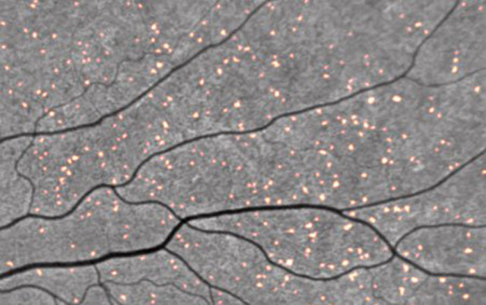
Amyloid deposits tagged by curcumin fluorescence in a retinal scan.
The researchers compared the number of retinal spots in a small subset of participants from each study. They found that spots of these proteins on the retina correlated with brain scans that showed higher levels of cerebral amyloid.
 Source/NeuroVision